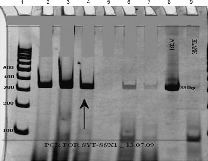
Fig. 4.

Polymerase chain reaction (PCR) analysis of SYT-SSX translocation using SYT and SSX1 primers. Reactions were subjected to electrophoresis on 10% polyacrylamide gel. Lane 1: the DNA size markers in base pairs (bp); Lanes 2 and 3: PCR run performed with cDNA from an already reported positive cases (331 bp) acting as positive control; Lane 4: PCR run performed with cDNA from test sample (arrow) showing positive band (331 bp); Lane 5: PCR run performed with cDNA from an already reported negative case acting as negative control; Lane 6: PCR run performed with cDNA from an earlier case, revealing weak band, interpreted as “inconclusive”; Lane 7: PCR run performed with cDNA from an unrelated tumour acting as negative control; Lane 8: Positive control DNA (pTZ57R/T-SYT-SSX1-331bp); Lane 9: PCR amplification without DNA template (Blank) to rule out contamination.
