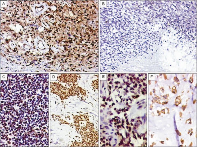
Fig. 5.

A. Diffuse TLE1 positivity in a neurilemoma/schwannoma. TLE1 positivity also noted within endothelial cells of vessels. DAB × 200. B. TLE1 negativity in MPNST (high-grade) DABx 200C. C. TLE1 positivity (2+) in a case of Ewing sarcoma/PNET. DAB × 200. D. TLE1 positivity (3+) in a desmoplastic small round cell tumour (DSRCT). DAB × 200. E. TLE1 positivity (2+) noted in a case of adamantinoma. DAB × 400. F. Negative nuclear staining, but positive cytoplasmic staining for TLE1 in chordoma. DAB × 400.
