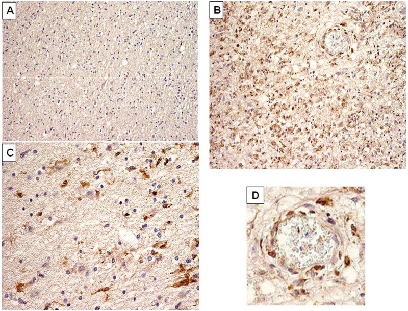
Figure 6.
Multiple sclerosis (MS) brain immunohistology evidencing Human Endogenous Retroviral family ‘W’ (HERV-W)/MS associated retroviral element (MSRV) envelope (Env) protein in MS brain lesions. (A) Normal appearing white matter (magnification ×10); (B) early active lesion with numerous Env-positive cells stained in brown (magnification ×40); (C) edge of active lesion, showing less numerous Env-positive cells stained in brown (magnification ×40); (D) detail of (B), magnifying a vascular element within the lesion with perivascular Env-positive cells stained in brown. These cells present morphological features and perivascular dissemination of macrophages. Immunohistological staining is representative of anti-HERV-W Env specific staining in different sections. Brown cells represent positive cells, significantly labelled by anti-Env monoclonals (examples with GN-mAb_03 are presented in this figure).

