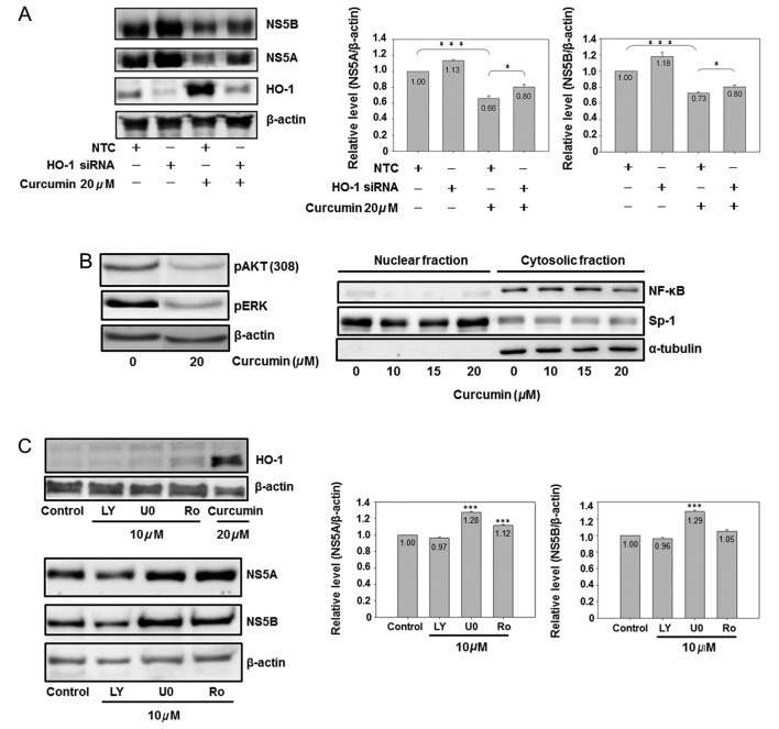
Figure 5.
The role of HO-1, AKT, ERK and NF-κB on curcumin-inhibited HCV protein expression is shown. (A) Knockdown of HO-1 partially reversed curcumin-inhibited HCV protein expression. Cells (3×10⁶) were seeded in a 10-cm dish for 6 h, and negative control small interfering (siRNA) (10 nM) or HO-1 siRNA (10 nM) was transfected into cells. Subsequent to a 6-h addition of siRNA, the medium was changed to fresh condition medium for 18 h, and the transfected cells were analyzed by western blotting (*P<0.05 and ***P<0.001, in 2 groups, respectively). (B) Curcumin inhibited AKT, ERK and NF-κB. Cells were subcultured at a density of 1.5×10⁶ cells in 8 ml of culture medium in a 10-cm plastic dish for 6 h. Curcumin or DMSO was added to the medium for 24 h. Total cell lysates (up) or cytosol-nuclear fraction (down) were isolated by western blot analysis. Sp1 is a dominant nuclear protein and α-tubulin is a cytosolic protein. (C) Effect of AKT, ERK and NF-κB inhibitors on the HCV protein expression is shown. Cells were subcultured at a density of 1.5×10⁶ cells in 8 ml of culture medium in a 10-cm plastic dish for 6 h. Chemical (LY, LY294002; U0, U0126; Ro, Ro1069920) or DMSO was added to the medium for 24 h. Total cell lysates were isolated for western blot analysis. (***P<0.001 compared to control). The experiments were repeated three times.

