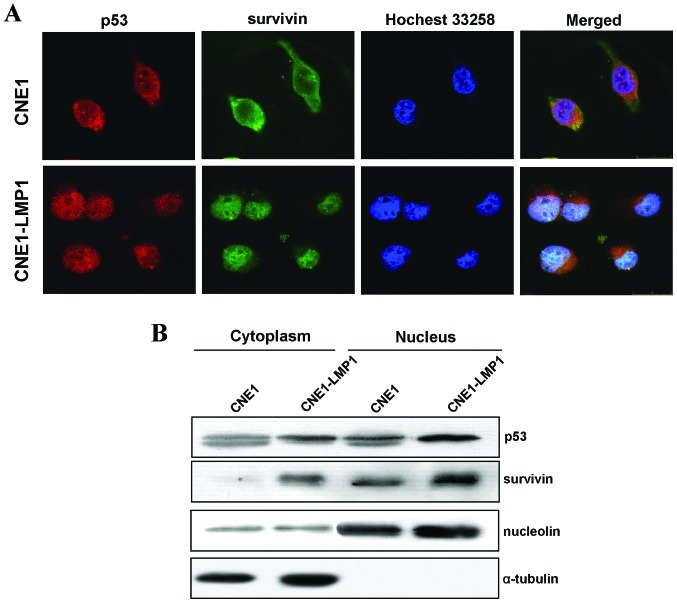
Figure 4.
(A) LMP1 promotes p53 and survivin nuclear accumulation, analyzed by immunofluorescence. Cy3 (red) staining indicates p53 localization, FITC (green) staining indicates survivin localization, and Hochest 33258 (blue) indicates nuclear staining. (B) Nuclear and cytoplasmic proteins were collected from CNE1 and CNE1-LMP1 cells and then separated by western blot analysis to detect survivin and p53. α-tubulin was used as a marker for cytoplasmic proteins and nucleolin for nuclear proteins.

