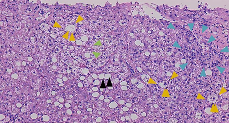
Fig. 3.
Pathological examination of liver biopsy (HE stain, ×100). The liver parenchyma shows a necrotic lesion accompanied by fibrosis and large droplets of fat (black arrowheads). The portal area was expanded due to infiltration of lymphocytes and fibrous tissue (blue arrowheads). No changes were seen in the bile ducts. Hepatocellular ballooning was evident (orange arrowheads). There was also mild to moderate fibrosis of the hepatic sinusoids (green arrowheads).

