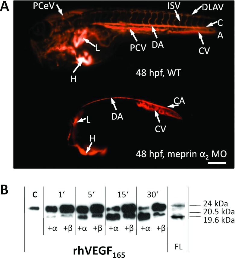
Figure 4. Meprin α promotes blood vessel formation.
To analyse the biological function of meprins, morpholino knockdowns of meprin α1, meprin α2 and meprin β were done in zebrafish embryos. (A) Microangiography with fluorigenic TRITC (tetramethylrhodamine β-isothiocyanate)–Dextran revealed severe defects in the formation of blood vessels when meprin α2 was reduced. Compared with wild-type (WT) embryos, meprin α2 morphants (MO) showed nearly complete absence of the head vascular system and the posterior cardinal vein (PCV), indicating a pro-angiogenic function of this protease. CA, caudal artery; CV, caudal vein; DA, dorsal aorta; DLAV, dorsal longitudinal anastomotic vessel; H, heart; hpf, hours post-fertilization; ISV, intersegmental vessels; L, liver; PCeV, posterior cerebral vein; PCV, posterior cardinal vein. Scale bar is 250 μm. Reproduced from [28], under the terms of the Creative Commons Attribution Licence. (B) Interestingly, processing of the VEGF-A (VEGF-A165) by meprins in vitro resulted in cleavage products comparable with those found in zebrafish lysates (FL). For detailed information see [28].

