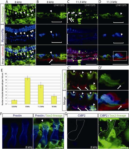
Figure 4. Lineage tracing of supporting cells in noise-exposed cochleae treated in vivo with a γ-secretase inhibitor.
(A) Double-labeled cells (arrowheads) positive for Sox2 lineage (GFP) and myosin VIIa (blue) were observed in the outer hair cell area (white bracket) in cochlear tissues from deafened mice carrying the Sox2-CreER as well as the Cre reporter transgene, mT/mG, 1 month after LY411575 treatment. Hair cell co-labeling with the lineage tag indicates derivation from a Sox2-positive cell and is thus evidence for regenerated hair cells after deafening in the mature mouse cochlea by transdifferentiation of supporting cells. These confocal xy-projection images of LY411575-treated ears from Sox2-CreER; mT/mG double transgenic mice are in the 8 kHz area of the cochlear longitudinal frequency map.
(B) Confocal xz-projections from the same area as A show that myosin VIIa-positive cells in the medial part of the outer hair cell area (white bracket) had GFP-positive hair bundle structures, indicating a Sox2 lineage (arrowhead). The cell shown was attached to the basement membrane (arrow) similar to a supporting cell.
(C) Cells double-labeled for myosin VIIa (blue) and Sox2 lineage (green) were observed (arrowheads) in the outer hair cell area (white bracket) in the 11.3 kHz region in this xy projection from a deafened cochlea 1 month after LY411575 treatment. Original hair cells have red hair bundles and new, Sox2-lineage hair cells have green (GFP-positive) bundles. (C') High power view of hair cells with their original (red) bundles (arrows) adjacent to cells with new (green) bundles (arrowheads) derived from Sox2-positive cells.
(D) Cross section from the same area as C shows that myosin VIIa, Sox2-lineage double-labeled cells in the outer hair cell area (white bracket) spanned the epithelium from the basement membrane to the endolymphatic surface.
(D') The cell shown is attached to the basement membrane (arrow) and its nucleus is at the base of the cell.
(E) Quantification of the GFP (Sox2 lineage) and myosin VIIa double-labeled cells in the outer hair cell region 1 month after treatment with LY411575 in deafened mice at frequency-specific cochlear areas (n = 5 in each group). Error bars are standard error of mean.
(F) Cells double-labeled for prestin (blue) and Sox2 lineage (green) were observed in the 8 kHz region in this xy projection from a deafened cochlea 1 month after LY411575 treatment. Sox2-lineage hair cell has green (GFP-positive) bundles (white arrowhead).
(G) Sox2-lineage hair cells (white broken line) were negative for CtBP2, which labels inner hair cell synaptic ribbons. White arrowhead indicates hair cell bundle.
A–D: Scale bars are 50 μm. F, G: Scale bars are 5 μm.

