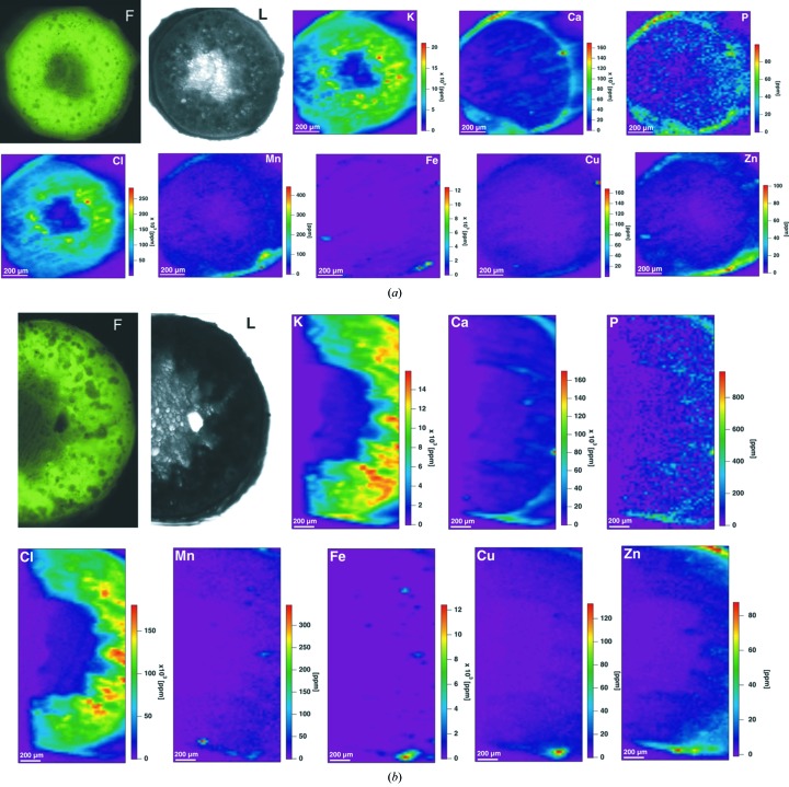
Figure 1.
X-ray fluorescence microscopy images of sections of the Dioscorea balcanica sixth internode, (a) twisted and (b) straight, at energy 13 keV. F: autofluorescence of the stem cross section; L: light microscopy of the investigated area. X-ray scanning mapping of K, Ca, P, Cl, Mn, Fe, Cu and Zn are shown. Scan areas: 1060 × 1450 µm (a) and 1320 × 1370 µm (b); pixel size: 15 µm; dwell time: 1 s.

