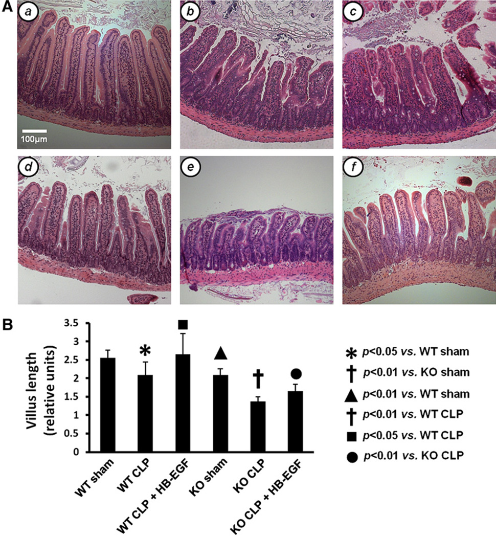
Fig 1.
Intestinal villous length. A, Representative H&E-stained histologic images of sections of jejunum from (a) WT mice subjected to sham operation (n = 5); (b) WT mice subjected to CLP (n = 4); (c) WT mice subjected to CLP but treated with HB-EGF (n = 6); (d) HB-EGF KO mice subjected to sham operation (n = 4); (e) HB-EGF KO mice subjected to CLP (n = 5); and (f) HB-EGF KO mice subjected to CLP but treated with HB-EGF (n = 5). Original magnification, ×100. B, Quantification of villous length. Villous length was measured as the distance from the crypt neck to the villous tip using Image J software. KO, HB-EGF KO mice; WT, wild type.

