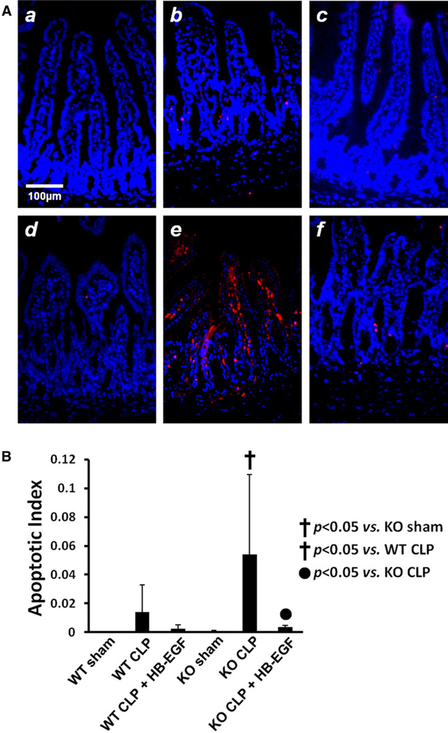
Fig 3.
A, Representative histologic images of sections of jejunum subjected to terminal deoxynucleotidyl transferase dUTP nick end labeling staining (red) and DAPI nuclear counterstaining (blue) from (a) WT mice subjected to sham operation (n = 5); (b) WT mice subjected to CLP (n = 9); (c) WT mice subjected to CLP but treated with HB-EGF (n = 7); (d) HB-EGF KO mice subjected to sham operation (n = 4); (e) HB-EGF KO mice subjected to CLP (n = 5); (f) HB-EGF KO mice subjected to CLP but treated with HB-EGF (n = 5). Original magnification, ×200. B, Positively stained enterocytes and total enterocytes were counted using Image Pro Plus 6.2.1 for Windows, and the apoptotic index was defined as the percent of positively stained enterocytes per 100 enterocytes. KO, HB-EGF KO mice; WT, wild type.

