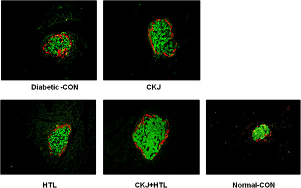
Figure 3.

Islet morphometry representing β-cells and α-cells. At the end of experiment, the α-cells were determined by immunostaining with a rabbit anti-glucagon in paraffin-embedded pancreatic section. Green and red were immunostained with anti-insulin and anti-glucagon, respectively and they are represented as β-cells and α-cells, respectively.
