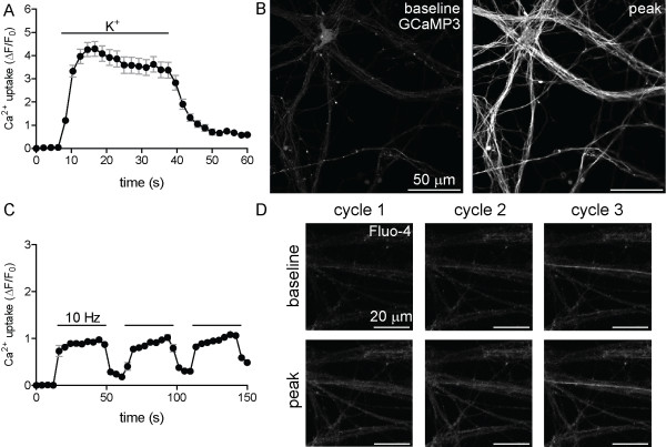
Figure 7.
Depolarization elicits reversible Ca2+ influx. Ca2+ uptake was evaluated following membrane depolarization by KEB (A, B) or three cycles of electrical field stimulation (10 Hz, 30 sec) (C, D). Time-lapse imaging of Ca2+ uptake (A, C) under depolarizing conditions, measured using the fluorescent reporter GCaMP3 (A; genetically encoded) or Fluo-4 (C). Solid black bars in A and C indicate the duration of treatment. Baseline and peak fluorescent intensities are demonstrated by the increase in GCaMP3 (B) or Fluo-4 (D) fluorescence. Error bars indicate standard error (A, C).

