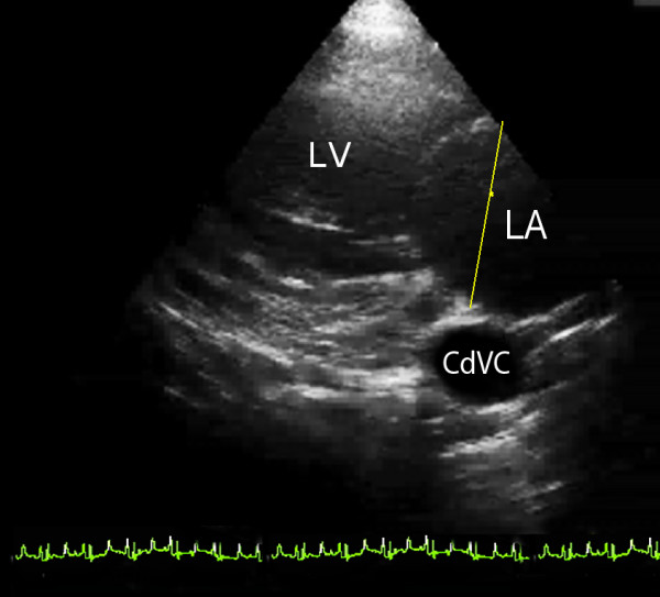
Figure 1.

Left parasternal caudal long axis view of the mitral valve, which is in the centre of the image. The left atrium (LA) is to the right of the image and the left ventricle (LV) to the left. The yellow line represents the position that the left atrium was measured. The caudal vena cava (CdVC) is used as a landmark for consistently obtaining this view.
