
Figure 1. Verification of p38β KO in microglia and brain.
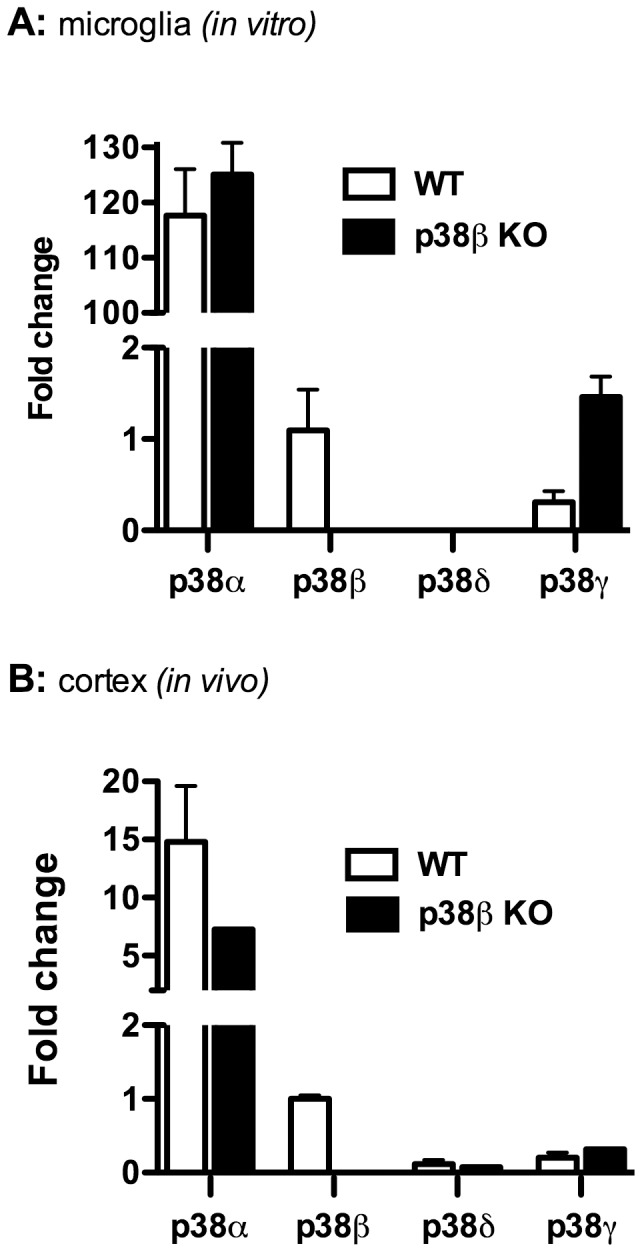
Primary microglia from mouse cortex were prepared as described in Methods and plated at 1×10⁴ cells/well in 48 well plates. Total RNA from microglia cultures (A) or from mouse cortical tissue (B) derived from WT (white bars) or p38β KO (black bars) mice was isolated, and the mRNA levels of different p38 MAPK isoforms were determined by qPCR. In both the microglia cultures and the brain tissues, p38β mRNA was readily measureable in WT mice but was not detected in the p38β KO mice. The p38α MAPK isoform in both microglia and cortex was expressed at much higher levels than any of the other isoforms, and there was no significant difference between the levels of p38α in either WT or p38β KO mice. The levels of p38δ mRNA were very low to undetectable in both WT and p38β KO mice. The expression of p38γ mRNA was slightly higher in microglia cultures from p38β KO mice compared to microglia from WT mice, but this difference was not seen in the cortical tissue samples. Results are expressed as fold change compared to p38β levels, and represent the mean ± SEM of two determinations.