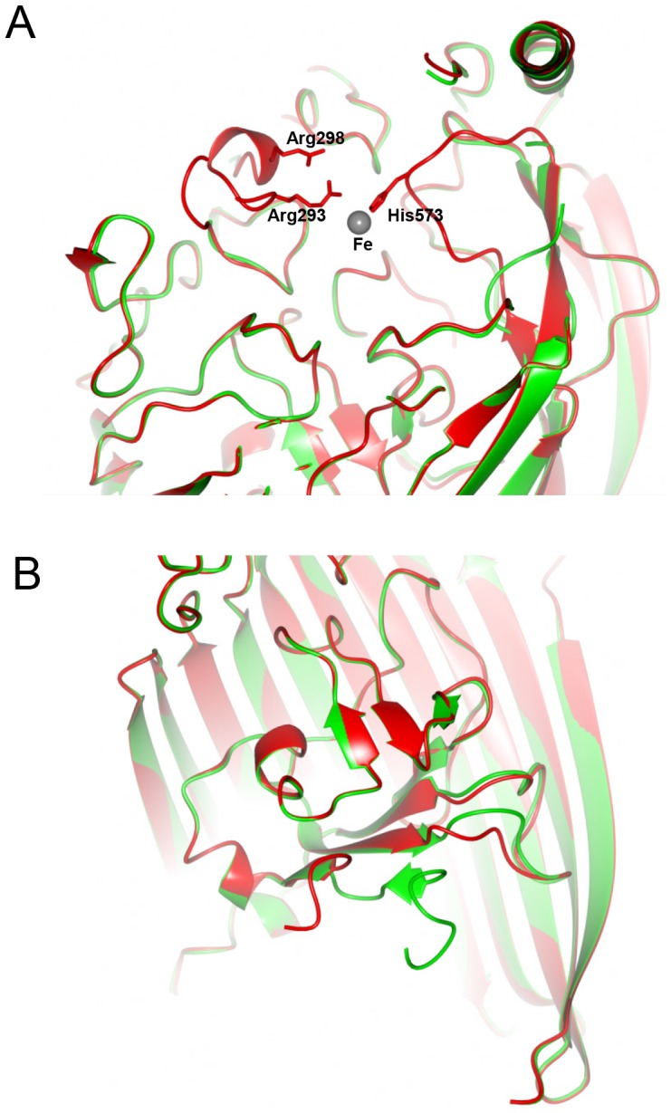
Figure 2. Structural changes on binding of Fe to FrpB F5-1.

(A) External loop movement during Fe recognition. The structure of FrpB F5-1 with Fe bound is shown in red, superimposed on the apoprotein form in green. The locations of Arg293, Arg298 and His573 are shown relative to the bound Fe atom. (B) Change in the conformation of the N-terminus as a result of Fe binding. Part of the β-barrel has been removed, for clarity. Colors used are as for part (A).
