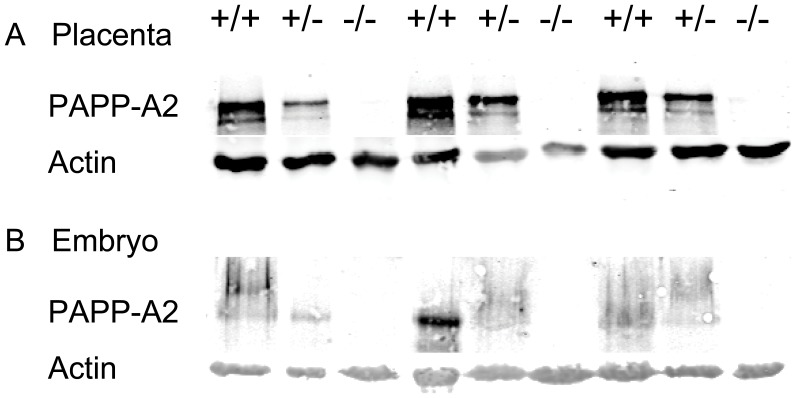
Figure 2. Effects of gene disruption on PAPP-A2 protein.
Shown are Western blots of 9 representative (A) placentae and (B) embryos at embryonic day 12.5 from embryos homozygous for wild-type Pappa2 (+/+), heterozygous (+/−) or homozygous for the disruption (−/−). The nitrocellulose membrane was scanned for fluorescence at 700 and 800 nm simultaneously, with fluorescence at 700 nm corresponding to actin (at approximately 40 kDa) and fluorescence at 800 nm corresponding to PAPP-A2 (at approximately 250 kDa).

