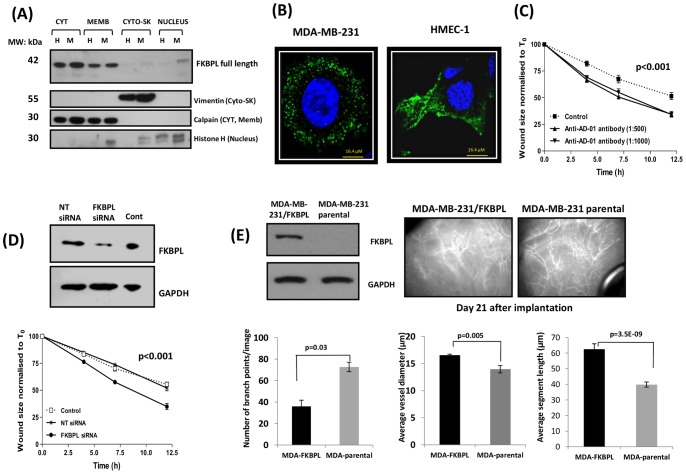
Figure 1. FKBPL is present in various cell compartments and regulates cell migration and tumour vasculature.
(A) Representative blot demonstrating that FKBPL is present predominantly in the cytosol and membrane compartments of both HMEC-1 (H) and MDA-MB-231 (M) cells and in the nuclear fraction of MDA-MB-231 cells. Protein extracts from each subcellular compartment probed with specific compartmental markers, vimentin, calpain and histone-H1 were used as loading controls. (B) Representative confocal images (60x) of MDA-MB-231 and HMEC-1 cells fixed, permeabilised and stained with DAPI (blue) and with anti-AD-01 primary antibody and Alexa-488 tagged secondary antibody demonstrating vesicular staining for FKBPL (green); n = 3. (C) Anti-AD-01 antibody targets the active domain of FKBPL and accelerates HMEC-1 cell migration in comparison to cells treated with an isotype control. Data points show means ± SEM; n = 3 (D) FKBPL knockdown with siRNA accelerated migration of HMEC-1 cells in comparison to un-transfected and NT-siRNA-transfected cells. Data points show means ± SEM; n = 3. Cell migration was assessed using scratch wound assay. Wound size is normalised to that of T0. p-value was determined using two-way ANOVA. (E) Intravital microscopy images (20x) representing disruption of tumour vasculature in vivo in FKBPL-overexpressing MDA-MB-231 xenografts in comparison to those derived from parental MDA-MB-231 cells. Tumours (21 days) were imaged using Epi-fluoresence microscopy following injection of mice with FITC-Dextran. Quantification of vessel dynamics was carried out on 3D images using ImageJ software. n = 5 mice per treatment group (p-value was determined using two-tailed T –test).

