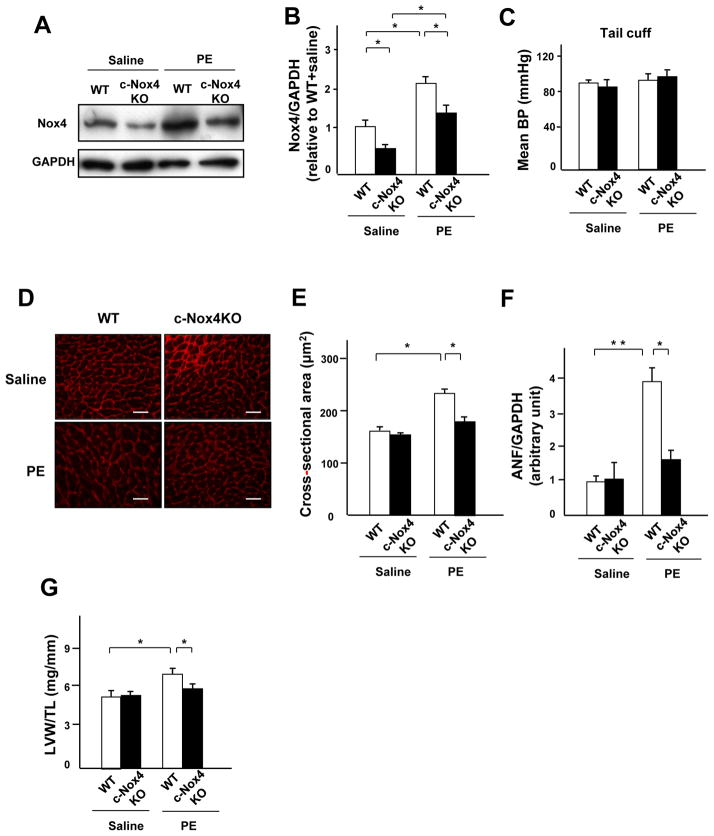
Figure 3. Endogenous Nox4 plays an essential role in mediating PE-induced cardiac hypertrophy.
A and B. Expression levels of Nox4 and GADPH in wild-type (WT) and c-Nox4 KO mouse hearts subjected to either PE or saline infusion for 2 weeks. The results of the quantitative analysis of Nox4 expression are shown (n=3). C. The effect of PE and knockdown of Nox4 on blood pressure was measured by tail cuff (n=6). D and E. LV myocyte cross-sectional area, as evaluated using WGA staining (n=4). Bar=20 μm. F. ANF mRNA normalized by GAPDH mRNA in WT and c-Nox4 KO mouse hearts in the presence or absence of PE infusion was evaluated by quantitative RT-PCR (n=6). G. LVW/TL after PE stimulation was determined (n=6). *P<0.05, **P<0.01.

