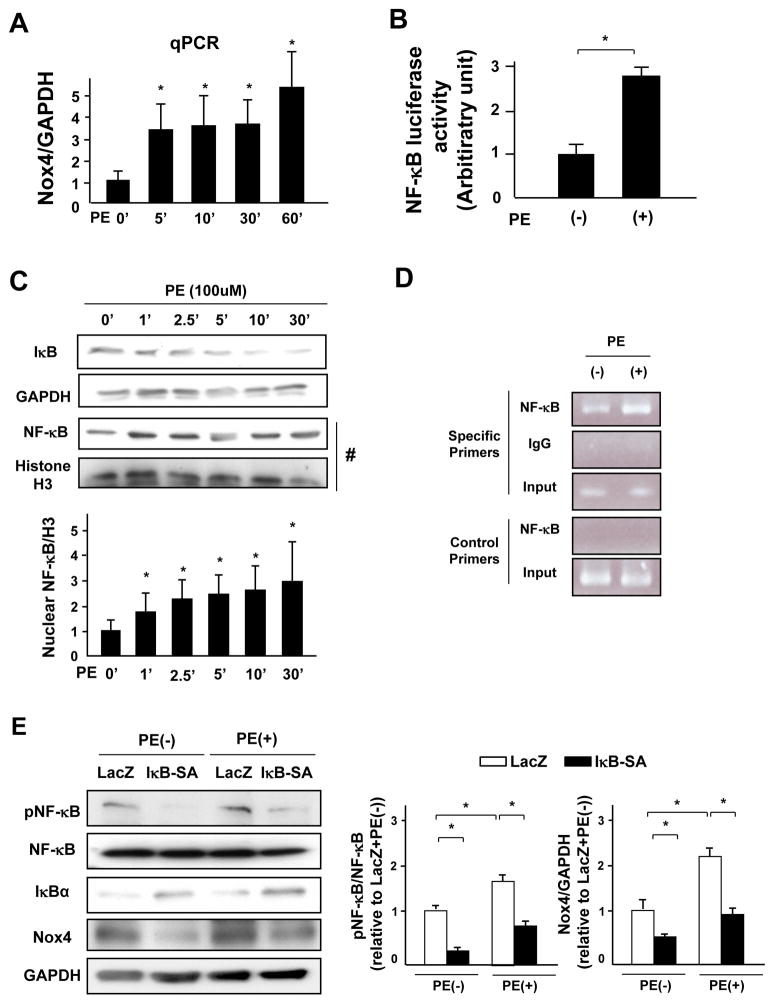
Figure 7. NF-κB mediates PE-induced Nox4 upregulation.
A. Myocytes were treated with PE for the indicated time periods. mRNA expression of Nox4 was determined by quantitative RT-PCR (n=5). B. Myocytes were treated with PE for 48 hours. NF-κB-luciferase activity was measured (n=8). C. Lysates were prepared from cardiomyocytes treated with PE for the indicated time periods. Protein expression of IκB and GAPDH was determined by immunoblotting. Nuclear fractions were prepared from myocytes treated with PE for the indicated time periods. Protein expression of NF-κB and histone H3 in the nuclear fraction was determined by immunoblotting (n=4). D. Chromatin immunoprecipitation (ChIP) assays with antibody against NF-κB (p65). A parallel ChIP assay was performed with rabbit IgG as an assay control. DNA was amplified and quantified by PCR with specific primers flanking the region of the rat Nox4 gene promoter containing the NF-κB-binding motif and with a pair of control primers that do not amplify the region containing the NF-κB-binding motif. PCR using input DNA as a template served as an internal control. The result shown is representative of 3 experiments. E. Myocytes transduced with the indicated adenoviruses were treated with PE for 48 hours. A representative analysis of the expression levels of phosphorylated NF-κB, NF-κB, IκB, Nox4 and GAPDH in the cultured neonatal rat cardiomyocytes and quantitative analyses of phosphorylated NF-κB/NF-κB and Nox4/GAPDH are shown (n=4). *P<0.05.

