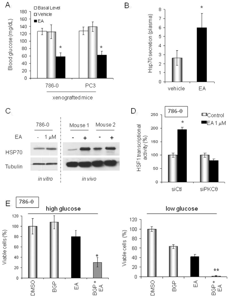
Figure 3. EA activates HSF1.
A. Effect of EA on blood glucose level in tumor-bearing mice prior to (“basal level”) or five minutes after treatment with EA (EA, 5mg/kg; vehicle: PBS/DMSO, 1/1). B. Quantification of plasma HSP70 level in tumor-bearing mice 4 h following EA treatment (EA: 5 mg/kg; vehicle: PBS/DMSO, 1/1). C. HSP70 protein expression following EA treatment of 786-0 cells in vitro (EA, 1 μM for 8 h) and in vivo (786-0 xenografts; prior to and 8 h after EA, 5mg/kg) was assessed by immunoblotting. D. EA stimulation of HSF1 transcriptional activity requires PKCθ expression. HSF1 activity was measured in cells transiently transfected with a HSP70 HSE-promoter GFP-tagged reporter plasmid 6 h after EA treatment. E. EA cytotoxicity is affected by HSF1 activation and extracellular glucose concentration. BGP-15 prolongs the HSF1 transcriptional response. High glucose medium contains 4.5g/L glucose; low glucose medium contains 1g/L glucose. BGP-15 was used at 50 μM (Chung et al., 2008) and EA was used at 1 μM. Incubation was for 24 h. Viability was assessed by MTT assay and confirmed by manual cell counting of trypan blue-excluding cells using a hemacytometer. *, p<0.05; **, p<0.001. Data are displayed as mean +/− SD. (see also Fig. S3)

