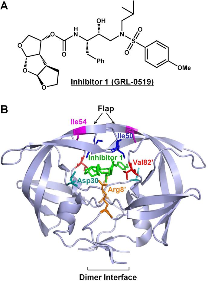
Figure 1.
(A) The chemical structures of protease inhibitor compound 1. (B) The structure of HIV-1 PRWT/inhibitor 1. The HIV-1 protease dimer is shown in light blue cartoon representation. The inhibitor 1 and wild-type residues at the mutation sites are indicated by differently colored sticks. The same residues on the two subunits are shown in the same color with only one of them labeled.

