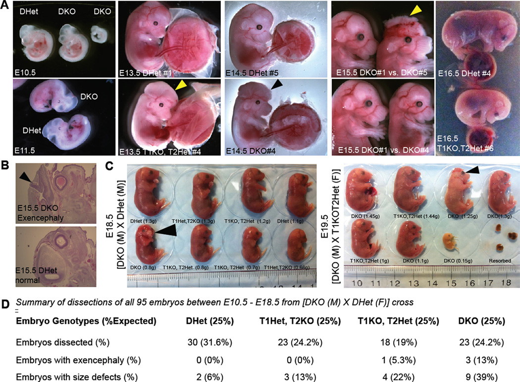
Figure 4. Defects in midgestation double knockout embryos.
(A) Representative images of mid-gestation E10.5 to E15.5 embryos of indicated genotypes. Notice the presence of normal, smaller size and exencephalic DKO embryos. Arrowheads point to exencephaly/neural tube defects. All embryos are from crosses of DKO male × Dhet female. (B) H&E staining for histological analysis of exencephaly. (C) Images, genotypes and weights of E18.5 and E19.5 litters from the indicated crosses. Arrowheads point to exencephaly/neural tube defects. (D) Summary of genotypes, phenotypes and Mendelian frequency of all mid and late gestation embryos.

