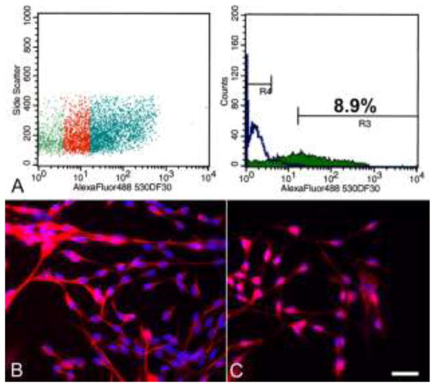
Fig. 4.
Canine CD15-positive cell population isolated from the rostral SVZ via flow cytometry. (A) The proportion of cells in the left panel that showed no overlap with the negative control (green region in left panel) are indicated in blue and comprise 8.9% of all gated cells (R3; right panel). The negative control cells are represented by R4 (right panel). Immunostaining of the CD15+ cells after one week in culture medium containing growth factors showed the cells are positive for nestin (B) and GFAP (C). Note the immature morphology of the cells and the diffuse, non-filamentous GFAP staining pattern. Bar = 25 μm.

