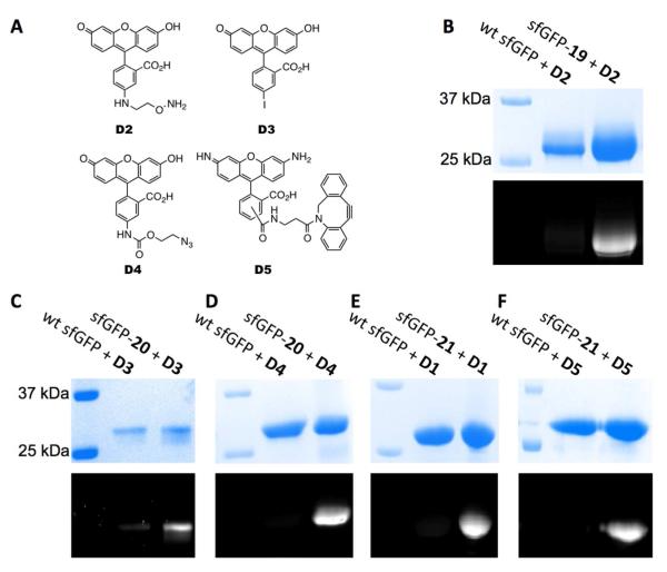
Figure 4.
(A) Structures of dyes D2-D5. (B) Specific labeling of sfGFP-19 with D2. (C) Specific labeling of sfGFP-20 with D3. (D) Specific labeling of sfGFP-20 with D4. (E) Specific labeling of sfGFP-21 with D1. (F) Specific labeling of sfGFP-21 with D5. In B-F, the top panels show denaturing SDS-PAGE analysis of sfGFP proteins with Coomassie blue staining and the bottom panels are from fluorescent imaging of the same gels before Coomassie blue staining. Wt sfGFP stands for wild-type sfGFP.

