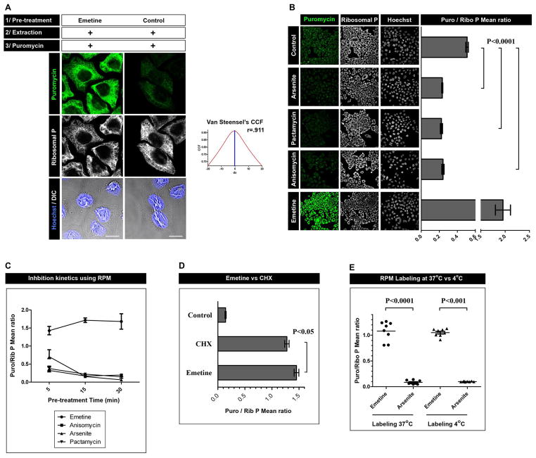
Figure 2.
A. HeLa cells were incubated or not for 15 min with emetine, were washed with PBS supplemented with CHX, permeabilized with digitonin and labeled with PMY in the presence of CHX. Cells were then fixed and stained for PMY (green) and large ribosomal subunit (white). Co-localization was quantitated using Van Steensel’s cross-correlation coefficient (graph) and Pearson’s coefficient (r; 1=complete co-localization, −1 = complete non-colocalization). Bar scale: 10 m.
B. HeLa cells pre-treated for 15 min with the inhibitor indicated were labeled as in A. For each condition, multiple fields were acquired and the mean fluorescence ratio of PMY/ribosome staining for each field was quantitated using ImageJ. Values are plotted on the right. Statistical analyses, two-tailed unpaired t-test; ***, P < 0.0001.
C. As in B, but with the treatment times indicated
D. As in B with CHX and emetine pre-treatments. Statistical analyses, two-tailed unpaired t-test; *, P < 0.05.
E. Direct comparison of RPM labeling using the 4 °C or 37 °C protocol. Statistical analyses, two-tailed unpaired t-test; ***, P < 0.0001.

