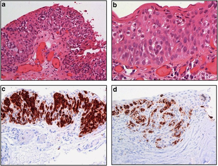
Figure 6.
Histological micrographs demonstrating invasive C-MIN. Epithelioid cells are present, some of which are arranged in nests. The tumour cells invade the substantia propria. Haematoxylin and eosin (a and b). The neoplastic cells are positive for melan-A (c and d). Courtesy of Sarah E Coupland.

