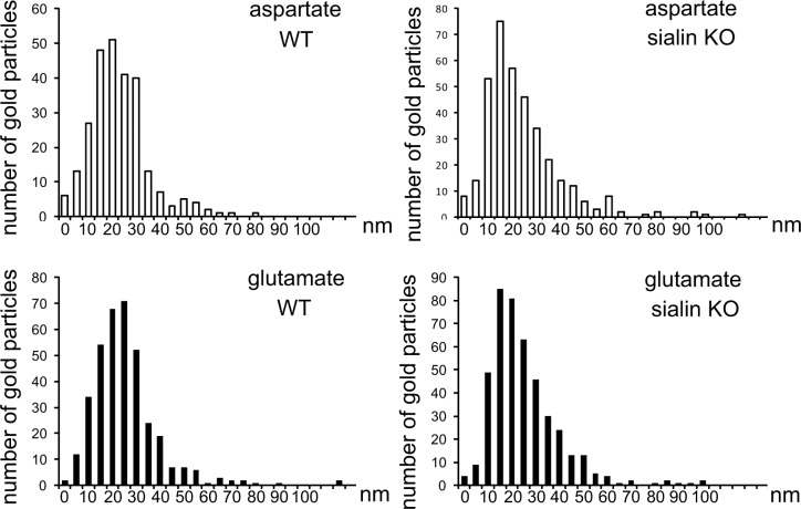
Figure 4.
l-Aspartate and l-glutamate are present in synaptic vesicles lacking sialin. Histograms show the frequency distribution of the distances separating l-aspartate and l-glutamate gold particles and synaptic vesicles in excitatory terminals in wild-type (WT) and sialin-knockout (KO) slices. Distances are put into bins of 5 nm (bars, x axis), and the number of gold particles in each bin is given along the y axis. Because of the lateral resolution of the immunogold method (∼30 nm), gold particles could be separated by a distance of up to ∼30 nm and still signal epitopes in the vesicle. Distributions of l-aspartate and l-glutamate intercenter distances in WT slices were not significantly different from the distributions in sialin-KO slices (P>0.05, χ2 test). These quantifications were done in 60 nerve terminals positive for aspartate/glutamate in both WT and sialin KO CA1 radiatum.

