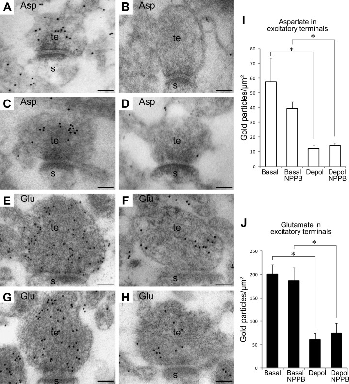
Figure 7.
l-Aspartate is not depleted from nerve terminals through volume-regulated anion channels. A–H) Electron micrographs show gold particles representing l-aspartate (A–D) or l-glutamate (E–H) in excitatory nerve terminals (t) making asymmetric synapses with dendritic spines (s) in the CA1 stratum radiatum. A, E) Terminals incubated at 3 mM K+. C, G) Terminals incubated at 3 mM K+ with NPPB. B, F) Terminals incubated at 55 mM K+. D, H) Terminals incubated at 55 mM K+ with NPPB. I, J) Quantification of gold particles representing l-aspartate (I) and l-glutamate (J) in excitatory nerve terminals in slices incubated at 3 mM K+ (basal), at 3 mM K+ with NPPB (basal NPPB) at 55 mM K+ (depol), and at 55 mM K+ with NPPB (depol NPPB). Bar charts indicate particle density (average±sem; gold particles/μm2). Quantifications in I and J were performed in 3 and 4 basal slices (total of 68 and 138 terminals, respectively), 3 basal NPPB slices (total of 132 and 91 terminals), 3–4 depol slices (total of 62 and 97 terminals), and 3–4 depol NPPB slices (total of 110 and 158 terminals). *P < 0.05; Student's 2-tailed t test (SPSS).

