
Figure 4.
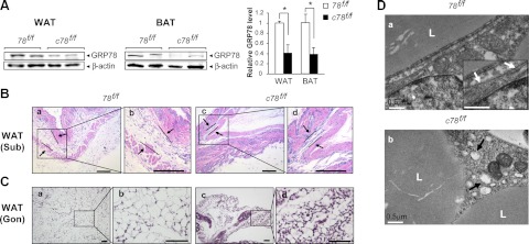
Residual white adipocytes from c78f/f mice display reduced lipid accumulation and dilated endoplasmic reticulum. A) Left panel: representative Western blot detection of GRP78 level in residual WAT and BAT isolated from 78f/f and c78f/f mice, with β-actin serving as loading control. Right panel: quantitation of GRP78 level after normalization to the β-actin level (n=3/genotype). Data are presented as means ± se. *P < 0.05. B) H&E staining of subcutaneous WAT tissues in 78f/f and c78f/f mice at postnatal d 23 at low magnification (a, c) and at high magnification (b, d) for the boxed regions. Arrows denote the boundaries of subcutaneous WAT. C) H&E staining of gonadal WAT from 78f/f and c78f/f mice at postnatal d 16 at low magnification (a, c) and at high magnification (b, d) for the boxed regions. D) Representative electron micrographs of white adipocytes from WAT in 78f/f mice (a) and c78f/f mice (b) at postnatal d 16. Inset (a): enlarged view showing normal ER structure, indicated by white arrows. Black arrows (b) indicate examples of expanded ER lumen in the adipocytes of c78f/f mice. L, lipid droplet. Scale bars = 200 μm (B); 50 μm (C); 0.5 μm (D).