
Figure 1.
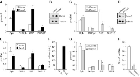
Spns2 transports S1P and dihydro-S1P from cells. A–D) Spns2 was overexpressed (A, B) or down-regulated (C, D) in HEK 293 cells. Then 48 h later, cells were washed, and culture medium was replaced with medium containing a phosphatase inhibitor. After 2 h, medium was collected and sphingosine (Sph), dihydrosphingosine (DHSph), S1P, and dihydro-S1P (DHS1P) were measured by LC-ESI-MS/MS (A, C). Data are means ± sd of triplicate determinations. *P < 0.05. B, D) In duplicate cultures, Spns2 expression was determined with anti-Spns2 antibody (Sigma-Aldrich). Blots were stripped and reprobed with anti-tubulin antibody to ensure equal loading and transfer. E–H) Similar results were found in 2 additional experiments. Spns2 was overexpressed (E, F) or down-regulated (G, H) in TIME cells. Sphingolipids were measured by LC-ESI-MS/MS (E, G), and mRNA levels of Spns2 were determined by quantitative real-time PCR and normalized to glyceraldehyde-3-phosphate dehydrogenase (F, H).