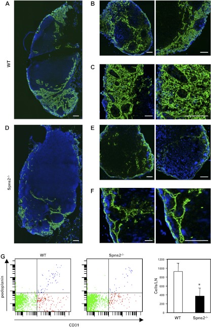
Figure 7.
Lack of Spns2 disrupts the lymphatic vessel network in lymph nodes. A–F) Immunofluorescent analysis of lymph nodes from wild-type (WT) littermates and Spns2−/− mice stained for LYVE-1 (green) and Hoechst (blue). Confocal images of axillary lymph nodes from wild-type (A–C) and Spns−/− mice (D–F), showing lymphatic sinus stained with antibody against LYVE-1 with low magnification using tile scan technology (A, B, D, E) and with high magnification (C, F). Scale bars = 100 μm. G) Single-cell suspensions were prepared from axillary lymph nodes of wild-type and Spns2−/− mice by digestion with collagenase, and total numbers of LECs were quantified by FACS analysis. Data are means ± se of triplicate determinations.

