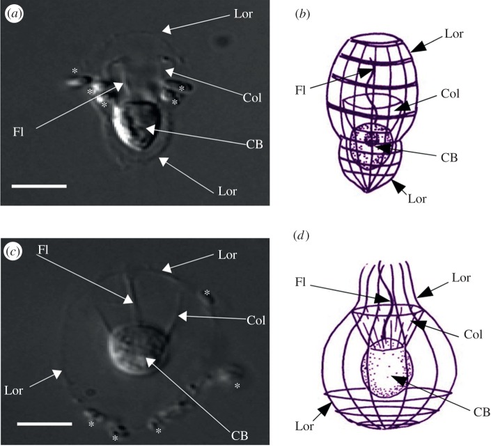
Figure 1.
The loricate choanoflagellates (a,b) S. diplocostata and (c,d) D. grandis. Photographs taken using phase contrast at 100× magnification. Schematic figures are taken from the Micro*scope v. 6.0 website (http://starcentral.mbl.edu/microscope, drawings by Won-Je Lee) and used under a Creative Commons licence. Lor, Lorica; CB, cell body; Fl, flagellum; Col, collar. Bacteria in the photographs are annotated with asterisks (*). Scale bar, 5 μm. (Online version in colour.)

