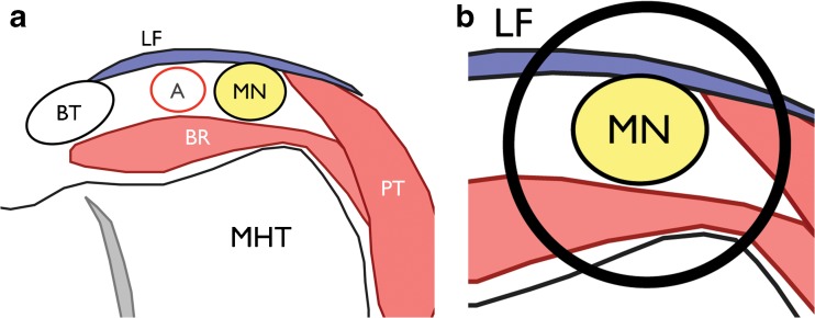
Fig. 3.
a Illustrations of a transverse section of the “lacertus tunnel” at the volar and medial aspect of the elbow, modified from Grant’s Atlas of Anatomy(2). The medial humeral trochlea (MHT) constitutes the bottom of the tunnel; the lateral and medial walls are the brachialis (BR) and pronator teres (PT) muscles, respectively, and the roof is the lacertus fibrosus (LF). MN = median nerve; A = brachial artery; BT = biceps tendon. b zoomed view of the lacertus tunnel

