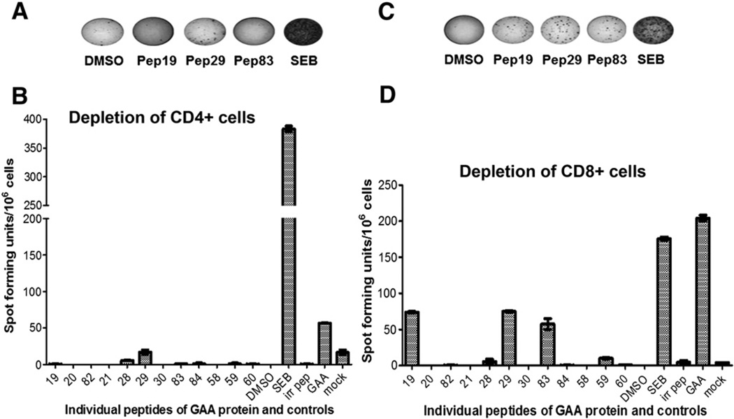
Fig. 4.
Confirmation that identified peptides contain CD4+ T cell epitopes. A. Representative examples of well images from IFN-γ ELISpot assays after stimulation of CD4+ cell-depleted splenocytes with peptides 19, 29, 83, rhGAA protein, SEB or solvent control. GAA−/− 129SVE had been immunized with rhGAA/CFA. B. Average frequencies of IFN-γ spot forming units (± SD, triplicate) after CD4+ depletion and stimulation with indicated peptides or controls. C. Representative examples of well images from IFN-γ ELISpot assays after stimulation of CD8+ cell-depleted splenocytes. D. Average frequencies of IFN-γ spot forming units (± SD, triplicate) after CD8+ depletion and stimulation with indicated peptides or controls.

