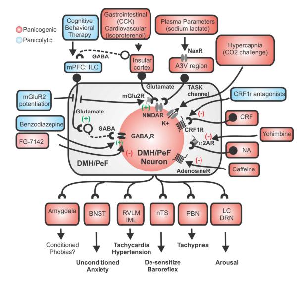
Figure 4.
Hypothetical schema of the sensory and neurochemical mechanisms by which panicogenic stimuli triggers panic-like responses in the hypothalamic panic model. Sources of GABA in the DMH/PeF that are inhibited by l-allyglycine include local interneurons, which are tonically driven by glutamatergic input from medial prefrontal cortex (mPFC) [41, 65]. Elevated plasma levels of Na+ from sodium lactate infusion are “sensed” by specialized NaX ion channels [101, 102] that are highly expressed in the anterior 3rd ventricle region (A3Vr) [103]. The AV3r contains many DMH/PeF projecting glutamatergic (Glu) neurons [87] which (following a hypertonic Na+ Challenge) excites the DMH/PeF region to mobilize panic-like responses. This figure also illustrates how other panic provoking stimuli such as hypercapnia [via CO2 sensitive K+ channels (TASK) [120]], cortocotrophin releasing factor (CRF) [89], isoproterenol, [211], cholecystokinin (CCK)[212], and yohimbine, caffeine and FG-7142 [213]] increase neuronal responses in the DMH/PeF. Afferent targets of the DMH/PeF which are implicated in the regulation of panic-like responses are also listed [see [68] CNS responses in panic model and also (Chamberlin and Saper, 1994;Chen et al., 2004;Fontes et al., 2001;Thompson and Swanson, 1998)]. GABAergic and glutamatergic neurons are represented by circles attached to dashed or solid lines, respectively. Abbreviations: BNST, bed nucleus of the stria terminalis; CBT, cognitive behavioral therapy; IML, intermediolateral cell column of spinal cord; LC, locus coeruleus; nTS, nucleus of solitary tract; PBN, parabrachial nucleus; RVLM, rostroventrolateral medulla.

