
Fig. 1. Hemodynamic and respiratory responses to hemorrhagic shock.
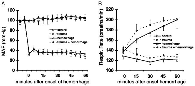
A–B, Changes in MAP (A) and respiratory rate before and during hemorrhagic shock (B). Control mice underwent catheter placement only. Trauma mice received 2-cm midline laparotomy and closure, followed by catheter placement. Hemorrhage mice underwent catheter placement, followed by hemorrhagic shock (blood withdrawal(s) necessary to maintain MAP = 35 ± 5 mmHg for 60 min). Trauma + hemorrhage mice received 2-cm midline laparotomy and closure, catheter placement, and hemorrhagic shock. Data are expressed as mean ± SD (n = 10 per group).