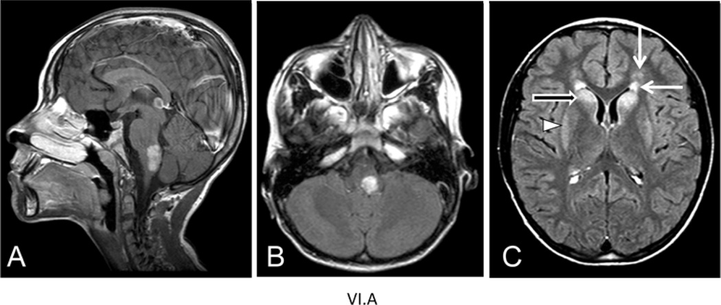
Figure 3.
MRI images from patient VI.A of family B at age 8.5 yrs. A) Sagittal T1-weighted image after contrast reveals an enhancing tumor-like lesion in the dorsal part of the medulla and lower pons. B) Axial FLAIR image shows the mass lesion in the medulla in the left posterior part. C) Axial FLAIR image shows signal abnormalities in the frontal white matter (closed arrows), head of the caudate nucleus (open arrow) and putamen (arrowhead).

