
Figure 8.
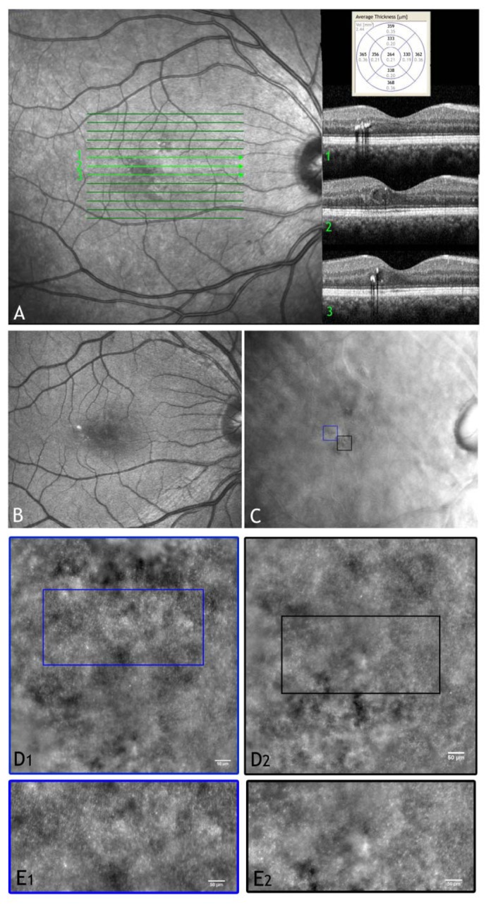
(A) Wide-field SLO image and OCT scans of the right eye in a 49 year old patient with mild non proliferative diabetic retinopathy showing hard exudates and a focal macular oedema. (B and C) Wide-field digital images (retromode modality by F10, Nidek, Japan) of the posterior pole showing the locations of the retinal exudate and the micro-cystic oedema (black box), respectively. (D1 and D2) Adaptive optics images of the photoreceptor layer acquired within the regions of interest enclosed in C (scale bars represent 50 μm). In panel D2, cones are highly resolved only in part probably due to increased scattering from oedematous inner retinal layers. (E1 and E2) High-magnification images of the photoreceptor layer shown in D1 and D2 respectively (scale bars: 50 μm). High variations in brightness between adjacent domains of photoreceptors can be seen in regions close to retinal oedema (E1). Intraretinal oedema reduces high-resolution imaging of photoreceptors (E2).