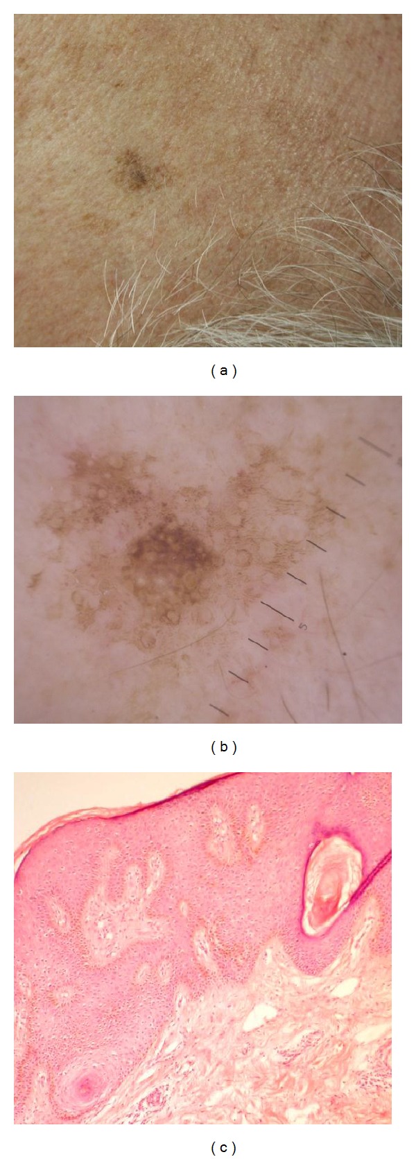
Figure 1.

(a) Seborrheic keratosis, temporal area (male, 67 years). (b) Same lesion dermoscopy: fissuring in the form of fingerprint-like structures around follicles, milia-like cysts, and comedo-like opening in the center of lesion. (c) Histopathology of seborrhoeic keratosis (acanthotic type) showing irregular epidermal hyperplasia mainly in the form of acanthosis (H&E ×200).
