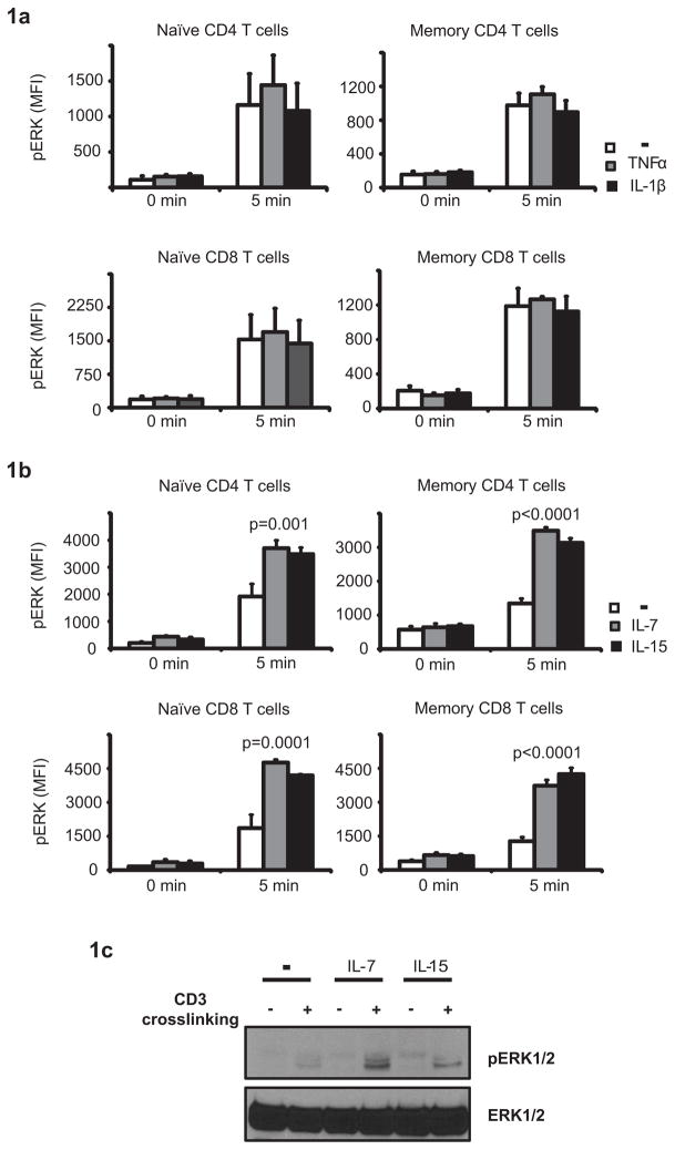
Figure 1. Cytokine-mediated priming of TCR-induced ERK signaling.
T cells were conditioned for 24 h with TNF-α, IL-1β (a), IL-7 or IL-15 (b and c). Cells were washed and stimulated by anti-CD3 crosslinking. Phosphorylation of ERK was analyzed for gated CD28+CD45RA+ CD4 and CD8 naïve and CD28+CD45RA− CD4 and CD8 memory T cells without and 5 min after TCR stimulation. Mean fluorescence intensities of pERK (MFI) are shown as mean ± SD of three to four experiments (b). Phosphorylation of ERK was confirmed using Western blot (c). The experiment shown is representative of three.

