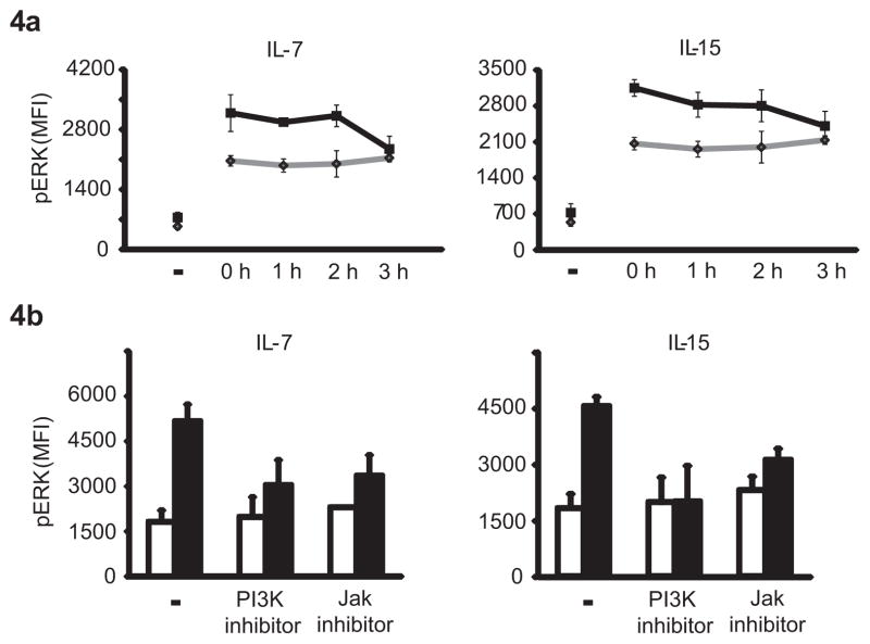
Figure 4. Cytokine-induced activation is mediated via the PI3K pathway and is short-lived.
(a) T cells conditioned with IL-7 or IL-15 for 24 h were washed and rested for 0–3 h prior to stimulation by crosslinking of CD3. Activation-induced ERK phosphorylation was measured after 5 min using PhosFlow. Results are shown as mean fluorescence intensities (MFIs) of pERK in T cells without (gray diamonds) or with prior cytokine exposure (black squares). (b) T cells were conditioned with IL-7 or IL-15 for 24 h in the presence or absence of 10 μM of the PI3K inhibitor LY294002 or 10 nM of pan JAK inhibitor. Cells were washed, stimulated by crosslinking CD3 and assessed for pERK. Results are shown as mean fluorescence intensities (MFIs) of pERK in T cells without (white bars) and with cytokine exposure (black bars). Data are shown as mean ± SD of T cells from three individuals.

