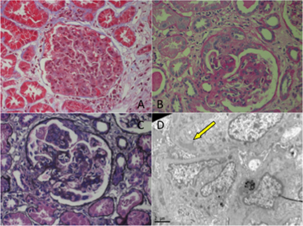
Figure 2.

A) Glomerular mesangial and endothelial cell proliferation (Masson’s trichrome; original magnification ×400).B). Small cellular crescent formation in several glomeruli (Periodic acid-Schiff; original magnification ×400). C) Subendothelial deposits (Masson’s trichrome and Jones methanamine silver; original magnification ×400). D) Glomerular hypercellularity with dense deposits (arrow) in the subendothelial area (electron microscopy; original magnification ×2000).
