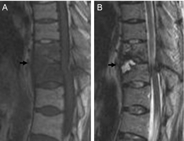
Figure 3.

Preoperative magnetic resonance imaging of a patient with Parkinson’s disease with a thoracic spine fracture in a severely ankylosed spine. The fracture site (T11: arrow) shows low intensity on the T1-weighted sagittal image (A) and high intensity on the T2-weighted sagittal image (B), suggesting the presence of fluid accumulation.
