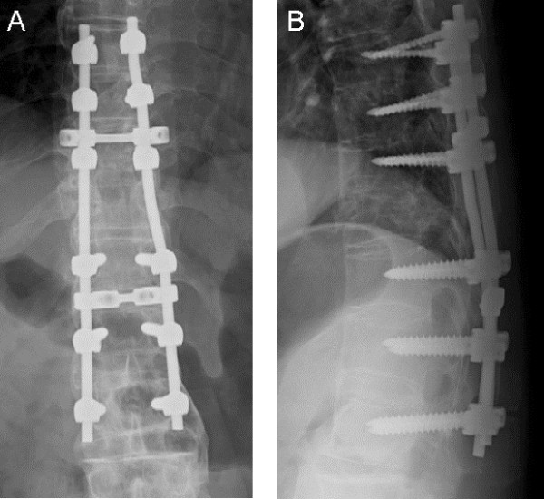
Figure 4.

Anteroposterior (A) and lateral (B) radiographs taken a week after surgery of a patient with Parkinson’s disease with a thoracic spine fracture in a severely ankylosed spine. Pedicle screws were inserted from T8 to L2 levels, except in the fractured T11 vertebral body.
