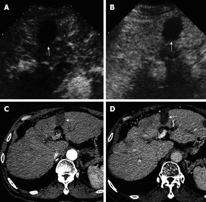
Figure 1.

A 57-year-old male patient with hepatocellular carcinoma. Two months after radiofrequency ablation for hepatocellular carcinoma in segment 3 of the liver. On both contrast-enhanced computed tomography (A, B) and contrast-enhanced ultrasound (C, D), the treated lesion (arrow) showed complete necrosis without any enhancement in all vascular phases.
