
Figure 4.
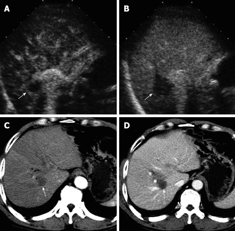
A 62-year-old male patient with hepatocellular carcinoma. Two months after percutaneous ethanol ablation for hepatocellular carcinoma in segment 8 of the liver. A, B: The false positive local tumor progression (arrow) was detected. It showed rim-like hyperenhancement in the arterial phase, wash-out in the portal-late phase on contrast-enhanced ultrasound; C, D: On contrast-enhanced computed tomography, the treated area (arrow) showed complete necrosis without any enhancement in all the vascular phases, but several enhanced hepatic vessels around the treated area.