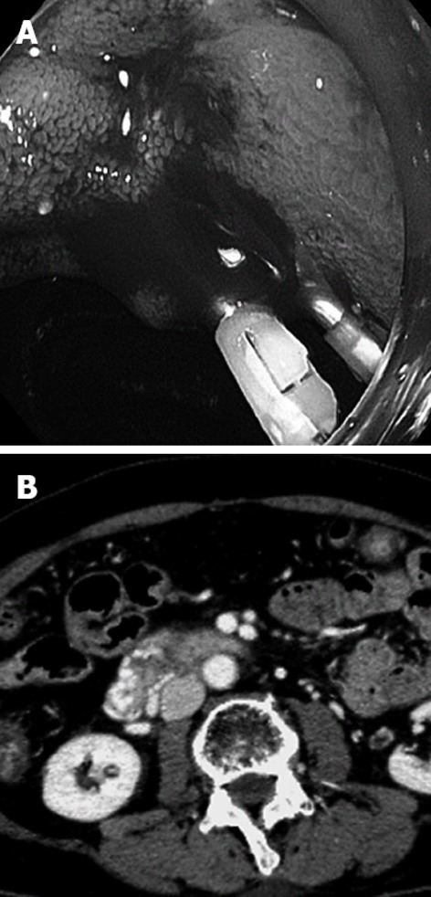
Figure 1.

Endoscopy and computed tomography of the duodenum. A: Endoscopy demonstrates bleeding varices in the second portion of the duodenum; B: Contrast-enhanced computed tomography reveals markedly tortuous varices around the wall in the second and third portion of the duodenum.
