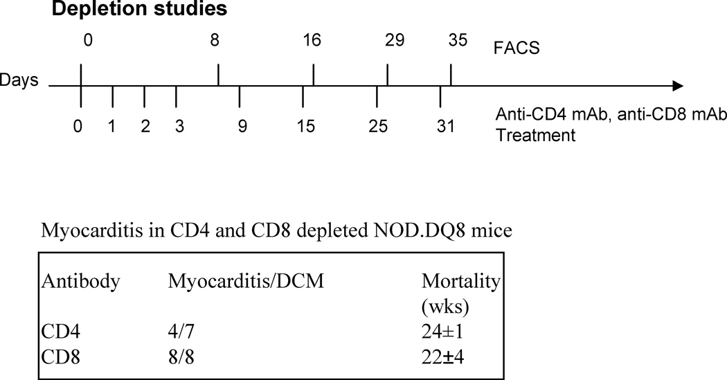
Figure 4.
Protocol for depleting CD4 and CD8 T cells in vivo. Mice were given intravenous injections of anti-CD4 (GK1.5) and anti-CD8 (lyt2) antibodies on indicated days. Presence of CD4 and CD8 cells in peripheral blood was analyzed by FACS after staining with specific conjugated antibodies on indicated days (top line). Most of the mice showed depletion of CD4 cells by 99% while CD8 T cells were depleted ranging between 85–95% in mice. Depleted mice were monitored for development of Myocarditis/dilated cardiomyopathy. CD4 depleted mice showed a later mortality as compared to nondepleted mice while CD8 depleted mice showed myocarditis in 100% of mice.

