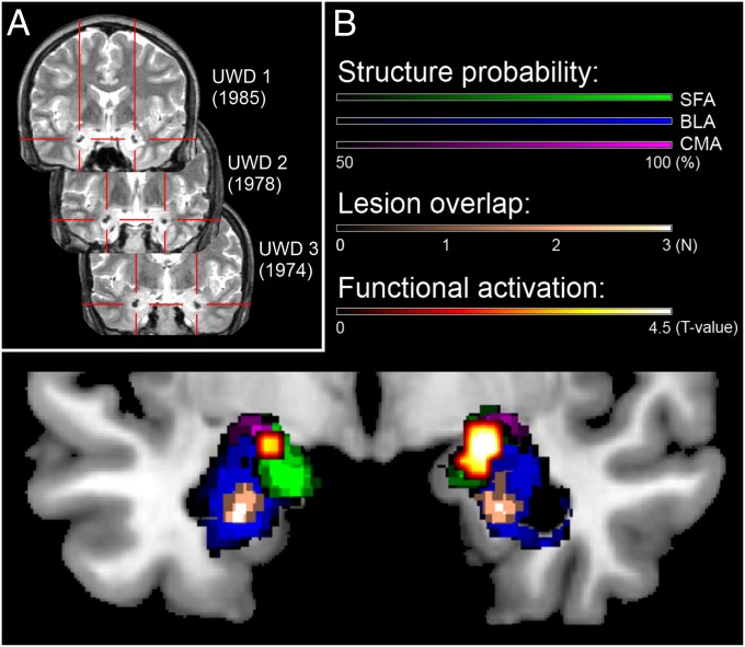
Fig. 1.
(A) T2-weighted MR images (coronal view) of the three subjects with Urbach–Wiethe disease (UWD), with their year of birth and red crosshairs indicating the calcified brain damage. (B) Structural and functional assessment of the bilateral amygdala in our group of three UWD subjects. Plotted are the cytoarchitectonic probability maps of the amygdala thresholded at 50% (47), structural lesion overlap, and functional activation during the emotion-matching task (48), all normalized to the Montreal Neurological Institute template brain. The structural method indicates that the lesions of the three patients are located in the BLA, whereas the functional method shows activation during emotion matching in the superficial amygdala (SFA) and CMA, but not in the BLA. This figure is adapted from Morgan et al. (38), wherein detailed structural MRI and functional MRI methods used are described.

